Cardiac Nuclear Medicine
Cardiac nuclear medicine imaging evaluates the heart for coronary artery disease and cardiomyopathy (diseases of the heart muscle). It also may be used to help determine whether the heart has been damaged by chemotherapy or radiotherapy. Nuclear medicine uses small amounts of radioactive materials called radiotracers that are typically injected into the bloodstream, inhaled or swallowed. The radiotracer travels through the area being examined and gives off energy in the form of gamma rays which are detected by a special camera and a computer to create images of the inside of your body. Nuclear medicine imaging provides unique information that often cannot be obtained using other imaging procedures.
Tell your doctor if there's a possibility you are pregnant or if you are breastfeeding and discuss any recent illnesses, medical conditions, allergies and medications you're taking. Depending on the type of exam, your doctor will instruct you on what you may eat or drink beforehand, especially if sedation is to be used. Leave jewelry at home and wear loose, comfortable clothing. You may be asked to wear a gown.
- What is Cardiac Nuclear Medicine?
- What are some common uses of the procedure?
- How should I prepare?
- What does the equipment look like?
- How does the procedure work?
- How is the procedure performed?
- What will I experience during and after the procedure?
- Who interprets the results and how do I get them?
- What are the benefits vs. risks?
- What are the limitations of Cardiac Nuclear Medicine?
What is Cardiac Nuclear Medicine?
Nuclear medicine uses small amounts of radioactive material called radiotracers. Doctors use nuclear medicine to diagnose, evaluate, and treat various diseases. These include cancer, heart disease, gastrointestinal, endocrine, or neurological disorders, and other conditions. Nuclear medicine exams pinpoint molecular activity. This gives them the potential to find disease in its earliest stages. They can also show whether you are responding to treatment.
Cardiac nuclear medicine is useful in diagnosing and assessing coronary artery disease. It is also used to evaluate cardiomyopathy and identify possible damage to the heart from chemotherapy or radiotherapy.
Nuclear medicine is noninvasive. Except for intravenous injections, it is usually painless. These tests use radioactive materials called radiopharmaceuticals or radiotracers to help diagnose and assess medical conditions.
Radiotracers are molecules linked to, or "labeled" with, a small amount of radioactive material. They accumulate in tumors or regions of inflammation. They can also bind to specific proteins in the body. The most common radiotracer is F-18 fluorodeoxyglucose (FDG), a molecule similar to glucose. Cancer cells are more metabolically active and may absorb glucose at a higher rate. This higher rate can be seen on PET scans. This allows your doctor to detect disease before it may be seen on other imaging tests. FDG is just one of many radiotracers in use or in development.
You will usually receive the radiotracer in an injection. Or you may swallow it or inhale it as a gas, depending on the exam. It accumulates in the area under examination. A special camera detects gamma ray emissions from the radiotracer. The camera and a computer produce pictures and supply molecular information.
Many imaging centers combine nuclear medicine images with computed tomography (CT) or magnetic resonance imaging (MRI) to produce special views. Doctors call this image fusion or co-registration. Image fusion allows the doctor to connect and interpret information from two different exams on one image. This leads to more precise information and a more exact diagnosis. Single photon emission CT/CT (SPECT/CT) and positron emission tomography/CT (PET/CT) units can perform both exams at the same time. PET/MRI is an emerging imaging technology. It is not currently available everywhere.
Cardiac nuclear medicine exams provide pictures of the distribution of blood flow to the heart muscle and can be used to visualize the function of the heart.
What are some common uses of the procedure?
Physicians use cardiac nuclear medicine studies to help diagnose cardiac disease. The symptoms include:
- unexplained chest pain.
- chest pain brought on by exercise (called angina).
- shortness of breath with exertion.
- abnormal electrocardiogram.
Cardiac nuclear medicine imaging is also performed:
- to visualize blood flow patterns to the heart walls, called a myocardial perfusion scan.
- to evaluate the presence and extent of suspected or known coronary artery disease.
- to determine the extent of injury to the heart following a heart attack, or myocardial infarction.
- to evaluate the results of bypass surgery or other revascularization procedures designed to restore blood supply to the heart.
- in conjunction with an electrocardiogram (ECG), to evaluate heart-wall movement and overall heart function with a technique called cardiac gating.
How should I prepare?
You may wear a gown during the exam or be allowed to wear your own clothing.
Women should always tell their doctor and technologist if they are pregnant or breastfeeding. See the Radiation Safety page for more information about pregnancy and breastfeeding related to nuclear medicine imaging.
Tell the doctor and your exam technologist about any medications you are taking, including vitamins and herbal supplements. List any allergies, recent illnesses, and other medical conditions.
You should inform your physician if you are pregnant or breastfeeding and/or if you have:
- had a recent heart attack or myocardial infarction
- heart failure
- asthma
- chronic lung disease
- conduction abnormalities within the heart (such as AV block), aortic stenosis or other abnormalities with the valves of your heart
- any abnormality with the heart and lungs
Also, if you have problems with your knees, hips or keeping your balance, tell your doctor as this may limit your ability to perform the exercise needed for this procedure. You should wear comfortable clothing and walking shoes. Do not apply oil, lotion, or cream to your skin the day of the exam. If you use an inhaler for asthma or other breathing problems, bring it to the test and make sure the health care team monitoring your stress test knows that you use an inhaler.
Leave jewelry and accessories at home or remove them prior to the exam. These objects may interfere with the procedure.
You should avoid caffeine (caffeinated as well as decaffeinated coffee, hot and cold tea, caffeinated soft drinks, energy drinks, chocolate and medications containing caffeine, etc.) and smoking for up to 48 hours before your examination. Your physician or radiologist may give you more specific instructions.
You should not eat or drink anything after midnight on the day of your procedure, but you may continue taking medications with small amounts of water unless your physician says otherwise. If you take beta-blocker or calcium channel blocker medication (Inderal, metoprolol, Norvasc, etc.) you should specifically ask your physician about temporary discontinuation. If you are diabetic, check with your physician about specific instructions for your diabetes medication on the day of the exam.
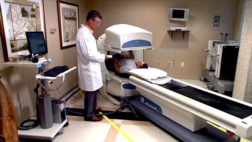
What does the equipment look like?
Nuclear medicine uses a special gamma camera and single-photon emission-computed tomography (SPECT) imaging techniques.
The gamma camera records the energy emissions from the radiotracer in your body and converts it into an image. The gamma camera itself does not emit any radiation. It has radiation detectors called gamma camera heads. These are encased in metal and plastic, often shaped like a box, and attached to a round, donut-shaped gantry. The patient lies on an exam table that slides in between two parallel gamma camera heads, above and beneath the patient. Sometimes, the doctor will orient the gamma camera heads at a 90-degree angle over the patient's body.
In SPECT, the gamma camera heads rotate around the patient's body to produce detailed, three-dimensional images.
Most nuclear medicine procedures use a gamma camera. Some nuclear medicine equipment has CT capabilities that help improve the images and increase the ability to combine functional imaging (nuclear medicine) and anatomic imaging (CT).
A computer creates the images using the data from the gamma camera.
How does the procedure work?
Ordinary x-ray exams pass x-rays through the body to create an image. Nuclear medicine uses radioactive materials called radiopharmaceuticals or radiotracers. Your doctor typically injects this material into your bloodstream. Or you may swallow it or inhale it as a gas. The material accumulates in the area under examination, where it gives off gamma rays. Special cameras detect this energy and, with the help of a computer, create pictures that detail how your organs and tissues look and function.
In order to evaluate the coronary arteries, heart scans are often performed immediately after patients have engaged in physical exercise (called a stress test) so that blood flow throughout the heart is maximized, making any blockages of the coronary arteries easier to detect. These images of the heart are compared with heart images taken while the patient is at rest. Patients who are unable to exercise are given a drug that increases blood flow to the heart.
How is the procedure performed?
Doctors perform nuclear medicine exams on outpatients and hospitalized patients.
You will be positioned on an examination table. A nurse or technologist will insert an intravenous (IV) line into a vein in your hand or arm.
The exam will usually begin with an injection of tracer while you are resting. Within the first hour after the tracer is injected, you will lie on a moveable imaging table with your arms (or in some cases your left arm only) over your head for about 15 to 20 minutes while images are recorded. Following imaging, you will undergo a stress test, which requires you to exercise either by walking on a treadmill or pedaling a stationary bicycle for a few minutes. While you exercise, the electrical activity of your heart will be monitored by electrocardiography (ECG) and your blood pressure will be frequently measured. When blood flow to the heart has reached its peak, you will be given the radiotracer through your IV. After you complete the stress test, you may be asked to drink some water. You will be placed on the imaging table a second time so a second series of images can be recorded. At this time, an ECG will also be placed to image the motion of your heart.
If you are unable to use a treadmill or bicycle, you will not exercise but you will be given a drug that will increase blood flow to the heart.
Actual scanning time for each heart scan varies from 15 to 30 minutes, depending on the type of scanner used. Total time in the nuclear medicine department will be approximately two to four hours.
If the SPECT scanner has CT capabilities, a short CT scan of your heart will be obtained. The CT may be obtained before or after each of the nuclear medicine imaging procedures. You will not have to get up or change positions on the table for this part of the exam as the CT is part of the nuclear medicine equipment, but you will be asked to stay very still for this portion of the exam.
After the exam, you may need to wait until the technologist determines if more images are needed. Sometimes, the technologist takes more images to clarify or better visualize certain areas or structures. The need for more images does not necessarily mean there was a problem with the exam or that something is abnormal. It should not cause you concern.
If you have an intravenous (IV) line for the procedure, your technologist will usually remove it. The technologist will leave it in place if you are to have another procedure that same day that requires an IV line.
What will I experience during and after the procedure?
Except for intravenous injections, most nuclear medicine procedures are painless. Reports of significant discomfort or side effects are rare.
You will feel a slight pin prick when the technologist inserts the needle into your vein for the intravenous line. You may feel a cold sensation moving up your arm during the radiotracer injection. Generally, there are no other side effects.
You will be asked to exercise until you are either too tired to continue or short of breath, or if you experience chest pain, leg pain, or other discomfort that causes you to want to stop.
If you are given a medication to increase blood flow because you are unable to exercise, the medication may induce a brief period of feeling anxious, dizzy, nauseous, shaky or short of breath. Mild chest discomfort may also occur. Any symptoms that do develop typically resolve as soon as the infusion is complete. In rare instances, if the side effects of the medication are severe or make you too uncomfortable, other drugs can be given to stop the effects.
It is important to remain still during the exam. Nuclear imaging causes no pain. However, having to remain still or in one position for long periods may cause discomfort.
Unless your doctor tells you otherwise, you may resume your normal activities after your exam. A technologist, nurse, or doctor will provide you with any necessary special instructions before you leave.
The small amount of radiotracer in your body will lose its radioactivity over time through the natural process of radioactive decay. It may also pass out of your body through your urine or stool during the first few hours or days after the test. Drink plenty of water to help flush the material out of your body.
Who interprets the results and how do I get them?
A radiologist or other doctor specially trained in nuclear medicine will interpret the images and send a report to your referring physician.
What are the benefits vs. risks?
Benefits
- Nuclear medicine exams provide unique information that is often unattainable using other imaging procedures. This information may include details on the function and anatomy of body structures.
- Nuclear medicine supplies the most useful diagnostic or treatment information for many diseases.
- A nuclear medicine scan is less expensive and may yield more precise information than exploratory surgery.
Risks
- If you have coronary artery disease, it is possible that you could experience chest pain during exercise or when a drug is given for the stress test. However, your heart will be monitored and if necessary, medication can be given for your chest pain.
- If life threatening cardiac disease is suspected because of the test findings, your cardiologist may consider same-day cardiovascular intervention.
- Because nuclear medicine exams use only a small dose of radiotracer, they have a relatively low radiation exposure. This is acceptable for diagnostic exams. Thus, the potential benefits of an exam outweigh the very low radiation risk.
- Doctors have been using nuclear medicine diagnostic procedures for more than six decades. There are no known long-term adverse effects from such low-dose exposure.
- Your doctor always weighs the benefits of nuclear medicine treatment against any risks. Your doctor will discuss the significant risks prior to treatment and give you an opportunity to ask questions.
- Allergic reactions to radiotracers are extremely rare and usually mild. Always tell the nuclear medicine personnel about any allergies you may have. Describe any problems you may have had during previous nuclear medicine exams.
- The radiotracer injection may cause slight pain and redness. This should rapidly resolve.
- Women should always tell their doctor and radiology technologist if there is any possibility that they are pregnant, or they are breastfeeding. See the Radiation Safety page for more information about pregnancy, breastfeeding and nuclear medicine exams.
What are the limitations of Cardiac Nuclear Medicine?
Nuclear medicine procedures can be time consuming. It can take several hours to days for the radiotracer to accumulate in the area of interest. Plus, imaging may take up to several hours to perform. In some cases, newer equipment can substantially shorten the procedure time.
The image resolution of nuclear medicine images may not be as high as that of CT or MRI. However, nuclear medicine scans are more sensitive for a variety of indications. The functional information they yield is often unobtainable using other imaging techniques.
This page was reviewed on March 11, 2024