Proton Therapy
Proton therapy delivers radiation to tumor tissue in a much more confined way than conventional photon therapy thus allowing the radiation oncologist to use a greater dose while still minimizing side effects.
If you're scheduled for proton therapy, you will likely undergo a treatment simulation session in which you will be fitted into an immobilization device to ensure you maintain the same exact position for every treatment. A CT scan will be performed to create a virtual model of your tumor, and other imaging procedures may be used to help determine its exact shape and location. Your doctor will give you specific instructions based on the type of exam being performed.
What is proton therapy and how is it used?
Protons are particles that carry a positive charge. Just as x-rays (also known as photons) are used to treat both benign and malignant tumors, protons beams can be used to irradiate tumors in a similar way. There is no significant difference in the biological effects of protons versus photons (x-rays). However, protons deliver a dose of radiation in a much more confined way to the tumor tissue than photons. After they enter the body, protons release most of their energy within the tumor region and, unlike photons, deliver only a minimal dose beyond the tumor boundaries. Therefore, especially for smaller tumor sizes, the dose of radiation may conform much tighter to the tumor and there may be less damage to healthy tissue. As a result, the treating physician (a radiation oncologist) can potentially give an even greater dose to the tumor while minimizing unwanted side effects. This is especially important when treating children, because protons help reduce radiation to growing and developing tissues.
Proton therapy is being used to treat tumors in these areas of the body with encouraging early results:
- Lung - see the Lung Cancer page
- Prostate - see the Prostate Cancer page
- Brain - see the Brain Tumors page
- Liver
- Breast
- Esophagus
- Rectum
- Skull base sarcomas
- Pediatric brain tumors
- Head and neck - see the Head and Neck Cancer page
- Eye melanomas
Protocols are being developed to explore the use of protons in other parts of the body.
Who will be involved in this procedure?
As with other forms of external beam therapy, proton beam therapy requires a treatment team, including a radiation oncologist, radiation physicist, dosimetrist, radiation therapist, and nurse. The radiation oncologist is a specially trained physician who evaluates the patient and determines the appropriate therapy, specific area for treatment, and radiation dose. Working together, the radiation oncologist, radiation physicist, dosimetrist and radiation therapist establish the best way to deliver the prescribed dose. Imaging exams are critical in delivering this treatment, and a diagnostic radiologist plays an important role as well. The radiation physicist and the dosimetrist make detailed treatment calculations to ensure treatment will be accurately delivered. Radiation therapists are specially trained technologists who perform the daily radiation treatments. Radiation therapy nurses are team members who tend to your day-to-day concerns and help to manage the side effects of the treatment.
What equipment is used?
Proton beam therapy uses special machines, a cyclotron and synchrotron being the most common, to generate and accelerate protons to speeds up to 60 percent the speed of light and energies of up to 250 million electron volts. These high-energy protons are steered by magnets toward the treatment room, and then to the specific part of the body being treated. In some older proton machines, additional pieces of equipment are needed to modify the range of the protons and the shape of the beam. Newer facilities make similar adjustments by fine tuning the energy of the beam and the magnetic fields which guide their path ("pencil beam scanning" or "scanning beam"). These modifications guide the proton beam to precise locations in the body where they deliver the energy needed to destroy tumor cells.
Who operates the equipment?
With backgrounds in mechanical, electrical, software, hardware and controls, specialized operators maintain, upgrade and repair the accelerators (i.e., cyclotrons and synchrotrons) and radiation delivery system. As with other forms of external beam radiation therapy, such as intensity-modulated radiation therapy (IMRT), specially trained radiation therapists will be present in the treatment rooms to help patients get set up for treatment, deliver the treatment, and monitor patients closely to ensure safety and comfort during the procedure. See the IMRT page for more information.
Is there any special preparation needed for the procedure?
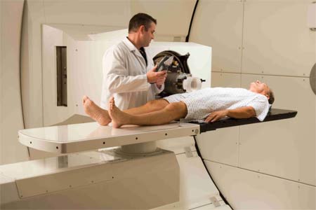
Several preparation steps are needed before a patient begins proton therapy. The first step in treatment preparation is a procedure referred to as "simulation," in which the patient is fitted into one of any number of specialized immobilization devices to ensure an exact, highly reproducible position for each treatment. Sometimes, there is a need for placement of a metallic marker (fiducial marker) in or around the tumor to help guide the treatment. If necessary, these procedures are usually performed a few days before the simulation.
The patient immobilization device used depends upon the location of the tumor. For treatments above the neck, a custom mask can be placed around the face, or custom tray that can be temporarily attached to the teeth. For treatments below the neck, custom molds of various shapes can be positioned under or around the body. Once the immobilization device is constructed, patients undergo a computed tomography (CT) scan, which is used to create a virtual, 3-D reconstruction of the tumor and normal tissues around it. Radiation oncologists define the boundaries of the tumor and the surrounding normal structures within this virtual computerized 3-D model, and work with physicists to determine the safest and most effective radiation techniques. This CT scan is performed in exactly the same position as for treatment, so the radiation oncologist can be sure the high-precision plan designed in the computer will accurately target the tumor.
Additional exams, such as a magnetic resonance imaging (MRI), or a PET/CT scan are often needed to help the physician accurately determine the location and extent of the tumor or critical structures.
How is the procedure performed?
Proton beam therapy is typically performed on an outpatient basis. For most tumor sites, a standard course of treatment is two to eight weeks, with treatments delivered five days per week. The length of each treatment will vary depending upon the tumor type and stage. The delivery of the proton beam to the patient lasts only a few minutes, although the total time spent in the treatment room will be longer (about 15 to 30 minutes) for positioning and adjustments to the equipment settings.
For daily treatments, the patient enters the treatment room and is fitted with his or her personal immobilization device. The patient is positioned with the aid of laser lights to within a few millimeters of the needed position, which by comparison with images obtained at the time of simulation to ensure the patient is properly aligned. In some cases, a CT system will be used to image the target before each treatment. This special alignment and imaging process is repeated before each treatment to assure the highest precision.
If special apertures and filters are needed for treatment, they are placed into position at some point during this process. A computer is typically used to scan and verify that the correct device has been loaded. Once positioning and treatment parameters are verified, the radiation oncologist and technologists step out into a control room located next to the treatment room and begin the treatment. During the treatment, these therapists continually monitor the progress of radiation delivery, including several safety parameters. Therapists also watch and listen to the patient closely, using video cameras, to ensure they remain safe and comfortable during the treatment. Once the prescribed radiation dose has been delivered, the computer shuts off the proton beam and the technologists re-enter the room to assist the patient in removing the mask or immobilization device.
What will I feel during and after the procedure?
You should not feel any pain or discomfort during the procedure. Afterward, there may be some side effects, and they will be managed by your radiation oncologist in the same way they would be for any course of radiation. Other factors that may influence how well you feel after treatment are how big a dose you are given and whether you are also getting chemotherapy at the same time. Common side effects include temporary hair loss and skin reactions in the direct path of the radiation and fatigue, especially when a large area is being treated.
Radiation treatment can cause side effects. These problems may result from the treatment itself or from radiation damage to healthy cells in the treatment area.
The number and severity of side effects will depend on the type of radiation, dose, and body part under treatment. Talk to your doctor and/or nurse so they can help manage them.
Radiation can cause early and late side effects. Early side effects happen during or right after treatment. They are typically gone within a few weeks. Common early side effects include fatigue and skin problems. Skin in the treatment area may become sensitive, red, irritated, or swollen. Other changes include dryness, itching, peeling, and blistering.
Depending on the area being treated, other early side effects may include:
- hair loss in the treatment area
- mouth problems and difficulty swallowing
- eating and digestion problems
- diarrhea
- nausea and vomiting
- headaches
- soreness and swelling in the treatment area
- urinary and bladder changes
Late side effects may occur months or years following treatment. While they are often permanent, they are rare. They include:
- brain changes
- spinal cord changes
- lung changes
- kidney changes
- colon and rectal changes
- infertility
- joint changes
- lymphedema
- mouth changes
- secondary cancer
There is a slight risk of developing cancer from radiation therapy. After treatment, your radiation oncologist will regularly check for complications and recurrent or new cancers.
The slight risk of developing cancer from radiation therapy is lower for proton than conventional x-ray (photon) treatment.
Highly advanced techniques such as proton therapy allow radiation oncologists to maximize the cancer-destroying capabilities of radiation, while minimizing its side effects.
This page was reviewed on March 11, 2024