

Image/Video Gallery
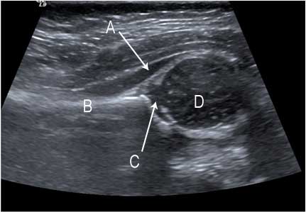
5 month old female: hip evaluated for congenital dysplasia
Ultrasound of a normal hip.
a. gluteus muscle
b. ilium
c. acetabulum
d. head of femur
Note: Images are shown for illustrative purposes. Do not attempt to draw conclusions or make diagnoses by comparing these images to other medical images, particularly your own. Only qualified physicians should interpret images; the radiologist is the physician expert trained in medical imaging.