Last reviewed on April 01, 2024
Abdominal and Pelvic CT
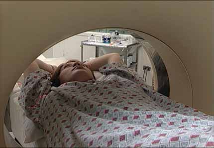
Computed tomography (CT) of the abdomen and pelvis is a diagnostic imaging test. There are many indications for a CT of your abdomen and pelvis. Your doctor may ask for this test when looking for kidney stones, other causes of abdominal pain or nausea / vomiting (including appendicitis and diverticulitis), and assessing a wide variety of cancers involving organs in your abdomen or pelvis. CT scanning is fast, painless, noninvasive and accurate. In emergency cases, it can reveal internal injuries and bleeding quickly enough to help save lives.
Tell your doctor if there's a possibility you are pregnant. Discuss any recent illnesses, medical conditions, medications you're taking, and allergies. Your doctor may tell you not to eat or drink anything for a few hours before your exam. If you have a known allergy to contrast material, your doctor may prescribe medications to reduce the risk of an allergic reaction. Leave jewelry at home and wear loose, comfortable clothing. You may be asked to change into a hospital gown for the exam.
What is CT Scanning of the Abdomen and Pelvis?
Computed tomography, more commonly known as a CT or CAT scan, is a diagnostic medical imaging test. Like traditional x-rays, it produces multiple images or pictures of the inside of the body.
A CT scan generates images that can be reformatted in multiple planes. It can even generate three-dimensional images.
CT images of internal organs, bones, soft tissue, and blood vessels provide greater detail than traditional x-rays. This is especially true for soft tissues and blood vessels.
What are some common uses of the procedure?
Doctors typically use this procedure to help diagnose the cause of abdominal or pelvic pain. They also use it to diagnose diseases of the internal organs, small bowel and colon, such as:
- infections such as appendicitis, diverticulitis, pyelonephritis or infected fluid collections, also known as abscesses.
- inflammatory bowel disease such as ulcerative colitis or Crohn's disease.
- pancreatitis.
- looking for or following cancers of the liver, bowel, kidneys, pancreas, adrenal gland, uterus/ovaries and bladder as well as lymphoma or melanoma. It is also commonly performed to look for spread of cancer into the abdomen/pelvis that form in other parts of the body such as lung cancer or breast cancer.
- kidney and bladder stones.
- abdominal aortic aneurysms (AAA).
- injuries to abdominal organs such as the spleen, liver, kidneys, or other internal organs in cases of trauma.
Doctors also use CT scanning of the abdomen/pelvis to:
- guide biopsies and other procedures such as abscess drainages and minimally invasive tumor treatments.
- plan for and assess the results of surgery, such as organ transplants.
- stage, plan and properly administer radiation treatments for tumors as well as monitor response to chemotherapy.
Who interprets the results and how do I get them?
A radiologist, a doctor specially trained to supervise and interpret radiology exams, will analyze the images. The radiologist will send an official report to the doctor who ordered the exam.
You may need a follow-up exam. If so, your doctor will explain why. Sometimes a follow-up exam further evaluates a potential issue with more views or a special imaging technique. It may also see if there has been any change in an issue over time. Follow-up exams are often the best way to see if treatment is working or if a problem needs attention.
What are the benefits vs. risks?
Benefits
- Viewing a CT scan, an experienced radiologist can diagnose many causes of abdominal pain or injury from trauma with very high accuracy. This allows for faster treatment and often eliminates the need for additional, more invasive diagnostic procedures.
- When pain is caused by infection and inflammation, the speed, ease and accuracy of a CT exam can reduce the risk of serious complications. Such complications may include those caused by a burst appendix or an infected fluid collection and the subsequent spread of infection.
- CT scanning is painless, noninvasive, and accurate.
- A major advantage of CT is its ability to image bone, soft tissue, and blood vessels all at the same time.
- Unlike conventional x-rays, CT scanning provides very detailed images of many types of tissue as well as the lungs, bones, and blood vessels.
- CT exams are fast and simple. In emergency cases, they can reveal internal injuries and bleeding quickly enough to help save lives.
- CT has been shown to be a cost-effective imaging tool for a wide range of clinical problems.
- CT is less sensitive to patient movement than MRI. While many patients have a hard time completing an MRI (because they take longer and can be more claustrophobic), most patients can tolerate a CT scan easily.
- Unlike MRI, an implanted medical device or metal of any kind in your body will not prevent you from having a CT scan.
- CT imaging provides real-time imaging, making it a good tool for guiding needle biopsies and needle aspirations. This is particularly true of procedures involving the lungs, abdomen, pelvis, and bones.
- A diagnosis via CT scan may eliminate the need for exploratory surgery and surgical biopsy.
- No radiation remains in a patient's body after a CT exam.
- The x-rays used for CT scanning should have no immediate side effects.
Risks
- There is always a slight chance of cancer from excessive exposure to radiation. However, the benefit of an accurate diagnosis far outweighs the risk involved with CT scanning.
- The radiation dose for this procedure varies. See the Radiation Dose page for more information.
- Women should always tell their doctor and x-ray or CT technologist if there is any chance they are pregnant. See the Radiation Safety page for more information about pregnancy and x-rays.
- The risk of serious allergic reaction to contrast materials that contain iodine is extremely rare, and radiology departments are well-equipped to deal with reactions.
- Doctors do not generally recommend abdomen and pelvis CT scanning for pregnant women unless medically necessary to avoid any radiation exposure to the baby. In general, there is no need to stop breast feeding after a CT scan.
- Because children are more sensitive to radiation, they should have a CT exam only if it is essential for making a diagnosis. They should not have repeated CT exams unless absolutely necessary. CT scans in children should always be done with low-dose technique.
- Radiology departments tailor the radiation dose for CT scans, especially when scanning children. This helps ensure that the benefits of the scan far outweigh any possible risks from exposure to diagnostic radiation.
What are the limitations of Abdominal and Pelvic CT?
A person who is very large may not fit into the opening of a conventional CT scanner. Or, they may be over the weight limit—usually 450 pounds—for the moving table.
CT scanning of the abdomen may not see gallstones well. Ultrasound is the best test for this.
Doctors prefer alternate imaging techniques such as plain films, gastrointestinal (GI) contrast exams and ultrasound for evaluating acute abdominal conditions in babies, such as vomiting or blood in stool.
For some conditions, including but not limited to some liver, kidney, pancreatic, uterine, or ovarian abnormalities, evaluation and diagnosis with MRI may be preferable to CT scanning.
Additional Information and Resources
RTAnswers.org: Radiation Therapy for Bladder Cancer
RTAnswers.org: Radiation Therapy for Colorectal Cancer