Last reviewed on July 15, 2026
Ultrasound - Vascular
Vascular ultrasound uses sound waves to evaluate the body's circulatory system and help identify blockages in the arteries and veins and detect blood clots. A Doppler ultrasound study – a technique that evaluates blood flow through a blood vessel – is usually part of this exam. Ultrasound does not use ionizing radiation, has no known harmful effects when used properly, and provides images of soft tissues that don't show up on x-ray images.
Little or no special preparation is required for this procedure. However, you may occasionally be asked to fast beforehand. Leave jewelry at home and wear loose, comfortable clothing. You may be asked to wear a gown.
What is Vascular Ultrasound?
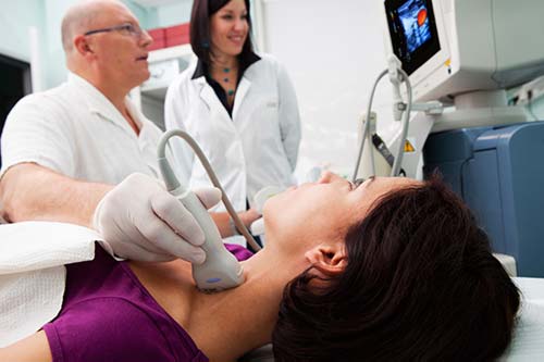
Ultrasound imaging is a noninvasive medical test that helps physicians diagnose and treat medical conditions. It is safe and painless. It produces pictures of the inside of the body using sound waves. Ultrasound imaging is also called sonography. It uses a small probe called a transducer and gel placed directly on the skin. High-frequency sound waves travel from the probe through the gel into the body. The probe collects the sounds that bounce back. A computer uses those sound waves to create an image. Ultrasound exams do not use radiation (x-rays). Because ultrasound captures images in real-time, it can show the structure and movement of the body's internal organs. The images can also show blood flowing through blood vessels.
Vascular ultrasound provides pictures of the body's veins and arteries.
A Doppler ultrasound study is usually part of a vascular ultrasound examination.
Doppler ultrasound is a special ultrasound technique that evaluates movement of materials in the body. It allows the doctor to see and evaluate blood flow through arteries and veins in the body.
What are some common uses of the procedure?
Sonography is a useful way of evaluating the body's circulatory system. Vascular ultrasound is performed to:
- help monitor the blood flow to organs and tissues throughout the body.
- locate and identify blockages (stenosis) and abnormalities like plaque or emboli and help plan for their effective treatment.
- detect blood clots (deep venous thrombosis (DVT) in the major veins of the legs or arms.
- determine whether a patient is a good candidate for a procedure such as angioplasty.
- evaluate the success of procedures that graft or bypass blood vessels.
- determine if there is an enlarged artery (aneurysm).
- evaluate varicose veins.
In children, ultrasound is used to:
- aid in the placement of a needle or catheter into a vein or artery to help avoid complications such as bleeding, nerve injury or pseudo-aneurysm (abnormal outpouching of an artery with the risk of rupture).
- evaluate a connection between an artery and a vein which can be seen in congenital vascular malformations (arteriovenous malformations or fistula) and in dialysis fistula.
If a line is placed in an artery or vein of the legs or arms, there is a much higher chance of developing a clot around it due to the smaller vessel size (especially in infants and young children). In some instances, a clot can form in the arm or in the left leg with the latter extending into major veins in the abdomen.
Doppler ultrasound helps the doctor to see and evaluate:
- blockages to blood flow (such as clots)
- narrowing of vessels
- tumors and congenital vascular malformations
- reduced or absent blood flow to various organs, such as the testes or ovary
- increased blood flow, which may be a sign of infection
Who interprets the results and how do I get them?
A radiologist, a doctor trained to supervise and interpret radiology exams, will analyze the images. The radiologist will send a signed report to the doctor who requested the exam. Your doctor will then share the results with you. In some cases, the radiologist may discuss results with you after the exam.
You may need a follow-up exam. If so, your doctor will explain why. Sometimes a follow-up exam further evaluates a potential issue with more views or a special imaging technique. It may also see if there has been any change in an issue over time. Follow-up exams are often the best way to see if treatment is working or if a problem needs attention.
What are the benefits vs. risks?
Benefits
- Most ultrasound scanning is noninvasive (no needles or injections).
- Occasionally, an ultrasound exam may be temporarily uncomfortable, but it should not be painful.
- Ultrasound is widely available, easy to use, and less expensive than most other imaging methods.
- Ultrasound imaging is extremely safe and does not use radiation.
- Ultrasound scanning gives a clear picture of soft tissues that do not show up well on x-ray images.
Risks
- Standard diagnostic ultrasound has no known harmful effects on humans when it is used properly by trained staff.
What are the limitations of Vascular Ultrasound?
- Vessels deep in the body are harder to see than superficial vessels. Specialized equipment or other tests such as CT or MRI may be necessary to properly visualize them.
- Smaller vessels are more difficult to image and evaluate than larger vessels.
- Calcifications that occur as a result of atherosclerosis may obstruct the ultrasound beam.
- The test is specialized and is best performed by a radiologic technologist and physician with experience in vascular ultrasound imaging.
- Image quality depends more on the skill and experience of the person performing the exam than it does with CT or MRI. Ultrasound is also less reliable for looking at the carotid artery where it runs deeper in the neck, and for telling a very narrow artery apart from one that is completely blocked. Your doctor may order a CT or MRI exam if the ultrasound cannot answer the question.
Additional Information and Resources
American Stroke Association: www.strokeassociation.org