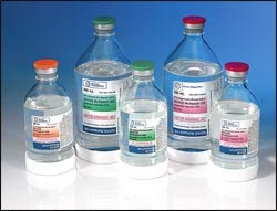
Contrast Materials
- What are contrast materials and how do they work?
- Which imaging exams use contrast materials?
- How safe are contrast materials?
- How should I prepare for my imaging procedure with contrast material?
- Side effects and adverse and allergic reactions
- What will I experience before and after receiving contrast material?
- Pregnancy and contrast materials
What are contrast materials and how do they work?
When a physician needs to understand what is happening inside our bodies, they often request that a patient undergo an imaging exam. Imaging exams such as x-rays, ultrasound, computed tomography (CT), magnetic resonance (MRI), and fluoroscopy are selected based on their ability to show specific information about the structures within the body. Contrast materials, also known as contrast agents or contrast media, are used to improve the diagnostic value of those imaging exams.
Contrast materials are not dyes that permanently discolor internal organs. They are substances that temporarily change the way x-rays or other imaging tools interact with the body. The materials discussed in this article do not produce radiation.
When introduced into the body prior to an imaging exam, contrast materials make certain structures or tissues in the body appear different on the images than they would if no contrast material had been administered. Contrast materials help distinguish or "contrast" selected areas of the body from surrounding tissue. This helps physicians diagnose medical conditions by improving the visibility of specific organs, blood vessels, or tissues.
Contrast materials enter the body in one of several ways. They can be:
- swallowed (taken by mouth or orally)
- administered by enema (given rectally)
- injected into a blood vessel (vein or artery; also referred to as being given intravenously or intra-arterially)
- injected into spaces within the body
Following an imaging exam with contrast material, the material is absorbed by the body or eliminated through urine or bowel movements.
There are several types of contrast materials:
- Iodine-based and barium-sulfate compounds are used in x-ray and computed tomography (CT) imaging exams.
Contrast materials can have a chemical structure that includes iodine, a naturally occurring chemical element. These contrast materials can be injected into veins or arteries, within the disks or the fluid spaces of the spine, and into other body cavities.
Barium-sulfate is the most common contrast material taken by mouth, or orally. It is also used rectally and is available in several forms, including:
- powder, which is mixed with water before administration
- liquid
- paste
- tablet
When iodine-based and barium-sulfate contrast materials are present in a specific area of the body, they block or limit the ability of x-rays to pass through. As a result, blood vessels, organs and other body tissue that temporarily contain the iodine-based or barium compounds change their appearance on x-ray or CT images.
- Gadolinium is the key component of the contrast material most often used in magnetic resonance (MR) exams. When this substance is present in the body, it alters the magnetic properties of nearby water molecules, which changes the appearance of the organs or blood vessels containing contrast when the MRI images are taken.
- Saline (salt water) and gas (such as air) are also used as contrast materials in imaging exams. Microbubbles and microspheres have been administered for ultrasound imaging exams, particularly exams of the heart.
Which imaging exams use contrast materials?
Oral Contrast Materials
Barium-sulfate contrast materials that are swallowed or administered by mouth (orally) are used to enhance standard x-ray, fluoroscopy, and CT images of the gastrointestinal (GI) tract, including:
- pharynx
- esophagus
- stomach
- the small intestine
- the large intestine (colon)
In some situations, iodine-based contrast materials are substituted for barium-sulfate contrast materials for oral administration.
Rectal Contrast Materials
Barium-sulfate contrast materials that are administered by enema (rectally) are used to enhance standard x-ray, fluoroscopy, and CT images of the lower gastrointestinal (GI) tract (colon and rectum). In some situations, iodine-based contrast materials are substituted for barium-sulfate contrast materials for rectal administration.
Intravenous Contrast Materials
Iodine-based and Gadolinium-based
Iodine-based contrast materials injected into a vein (intravenously) are used to enhance x-ray (including fluoroscopic images) and CT images. Iodine based contrast materials are also commonly injected in the arteries during angiogram procedures. Gadolinium injected into a vein (intravenously) is used to enhance MR images. Typically, these are used to enhance:
- internal organs, including the brain, breasts, heart, lungs, liver, adrenal glands, kidneys, pancreas, gallbladder, spleen, uterus, and bladder
- gastrointestinal tract, including the stomach, small intestine and large intestine
- arteries and veins of the body, including vessels in the brain, neck, chest, abdomen, pelvis and legs
- other parts of the body including muscle and bone
Microbubble Contrast Materials
Microbubble contrast materials are tiny bubbles of an injectable gas held in a supporting shell. They are extremely small—smaller than a red blood cell—and have a high degree of "echogenicity", or ability to reflect ultrasound waves. Structures with higher echogenicity will appear brighter on ultrasound. Once the microbubbles are in the bloodstream, ultrasound technology is able capture differences in echogenicity between the gas in the microbubbles and the surrounding tissues of the body, producing an ultrasound image with increased contrast. The microbubbles dissolve, usually within 10 to 15 minutes, and the gas within them is removed from the body through exhalation. Contrast-enhanced ultrasound with microbubbles is a convenient, relatively inexpensive way to improve visualization of blood flow, and it does not use radiation. It is a useful option for patients with kidney failure or those with allergies to contrast agents used for MR and/or CT imaging.
Microbubble contrast materials can be targeted or untargeted. Untargeted contrast-enhanced ultrasound —the more common method— helps diagnose certain diseases by providing evaluation of blood flow in the heart and other organs. In targeted contrast-enhanced ultrasound, specific molecules are bound to the surface of the microbubbles. After injection, the microbubbles attach to specific targeted tissue sites, causing an increase in the ultrasound signal at the sites.
Contrast-enhanced ultrasound with microbubbles is used in the assessment of:
- blood perfusion in organs
- thrombosis, such as in myocardial infarction
- abnormalities in the heart
- liver and kidney masses
- inflammatory activity in inflammatory bowel disease
- chemotherapy treatment response
How safe are contrast materials?
Contrast materials are safe drugs; adverse reactions ranging from mild to severe do occur, but severe reactions are very uncommon. While serious allergic or other reactions to contrast materials are rare, radiology departments are well-equipped to deal with them.
How should I prepare for my imaging procedure with contrast material?
Before arriving for you exam, you will be given specific instructions on how to prepare for the exam. Because contrast materials carry a slight risk of causing an allergic reaction or adverse reaction, you should tell your doctor about any of the following conditions. These conditions could affect the instructions you are given.
- previous allergic reactions to contrast materials
- allergies to food, drugs, dyes, preservatives, or animals
- history of heart disease, diabetes, kidney disease, or thyroid problems
Side effects and adverse and allergic reactions
Barium Sulfate Contrast Materials
You should tell your doctor if these mild side effects of barium-sulfate contrast materials become severe or do not go away:
- stomach cramps
- diarrhea
- nausea
- vomiting
- constipation
Tell your doctor immediately about any of these symptoms:
- hives
- itching
- red skin
- swelling of the throat
- difficulty breathing or swallowing
- hoarseness
- fast heartbeat
- bluish skin color
You are at greater risk of an adverse reaction to barium-sulfate contrast materials if:
- you have cystic fibrosis, which can increase the risk of blockage in the small bowel.
- you are severely dehydrated, which may cause severe constipation.
- you have an intestinal blockage or perforation that could made worse by a barium-sulfate agent.
Iodine-based Contrast Materials
A very small percentage of patients may develop a delayed reaction with a rash which can occur hours to days after an imaging exam with an iodine-based contrast material. Most are mild, but severe rashes may require medication after discussion with your physician
You should tell your doctor if these mild or moderate side effects of iodine-based contrast materials become severe or do not go away:
Mild reactions include:
- nausea and vomiting
- headache
- itching
- flushing
- mild skin rash or hives
Moderate reactions include:
- severe skin rash or hives
- wheezing
- abnormal heart rhythms
- high or low blood pressure
- shortness of breath or difficulty breathing
Tell your doctor immediately about any of these symptoms:
Severe reactions include:
- difficulty breathing
- swelling of the throat or other parts of the body
- profound low blood pressure
Contrast-Induced Acute Kidney Injury
Patients with impaired kidney (renal) function should be given special consideration before receiving iodine-based contrast materials by vein or artery. While many contrast agents are safe to give in patients with kidney disease, if you have severe kidney disease and very poor kidney function you may be at increased risk of worsening kidney function when getting iodinated contrast agents. The benefits of having a contrast enhanced scan often out-weigh the risks in ensuring the radiologist can properly diagnose your medical conditions.
MR-Gadolinium
The contrast material used in MRI (Magnetic Resonance Imaging) called gadolinium is less likely to produce an allergic reaction than the iodine-based materials used for x-rays and CT scanning. Very rarely, patients are allergic to gadolinium-based contrast materials and experience hives and itchy eyes. Reactions are usually mild and easily controlled by medication. Severe reactions are rare.
Nephrogenic systemic fibrosis (NSF), a thickening of the skin, organs, and other tissues, is a rare complication in patients with kidney disease that undergo an MR with contrast material. Gadolinium-based contrast material may be withheld in some patients with severe kidney disease.
There is evidence that tiny traces of gadolinium may be retained in different organs of the body, including the brain, after contrast-enhanced MRI. While there are no known negative effects from this, your doctor may take gadolinium retention into account when selecting a contrast agent. There are many different gadolinium-based contrast agents available, each with its own safety profile. Decisions on which material to use may be affected by the part of the body being imaged, the cost of the material and other factors. These decisions are especially important in patients likely to undergo multiple MRI scans with gadolinium-based contrast material, such as pediatric patients, cancer patients and people with multiple sclerosis.
What will I experience before and after receiving contrast material?
Barium-Sulfate Oral and Rectal Contrast Material
If a barium-sulfate contrast material (given orally or rectally) will be used during your exam, you may be asked not to eat for a few hours before your exam begins. If the contrast material will be given rectally, you may also be asked to cleanse your colon with a special diet and medication (possibly including an enema) before your exam.
If you swallow the contrast material, you may find the taste mildly unpleasant; however, most patients can easily tolerate it.
If your contrast material is given by enema, you can expect to experience a sense of abdominal fullness and an increasing need to expel the liquid. The mild discomfort will not last long.
It is a good idea to increase your fluid intake after an imaging exam involving a barium-based contrast material to help remove the contrast material from your body.
Barium-sulfate contrast materials are expelled from the body with feces. You can expect bowel movements to be white for a few days. Some patients may experience changes in their normal bowel movement patterns for the first 12 to 24 hours.
Iodine-based Contrast Material
When an iodine-based contrast material is injected into your bloodstream, you may have a warm, flushed sensation and a metallic taste in your mouth that lasts for a few minutes.
The needle may cause you some discomfort when it is inserted. Once it is removed, you may experience some bruising.
It is a good idea to increase your fluid intake after an imaging exam involving an iodine-based contrast material to help remove the contrast material from your body.
Gadolinium-based Contrast Material
When the gadolinium is injected, it is normal to feel coolness at the site of injection, usually the arm for a minute or two.
The needle may cause you some discomfort when it is inserted. Once it is removed, you may experience some bruising.
For all the above administrations of contrast material (barium sulfate, iodine-based, and gadolinium-based):
If you have not been sedated, no recovery period is necessary. You may resume your usual activities and normal diet immediately after the exam. Increased fluid intake will help eliminate the contrast material from your body.
Pregnancy and contrast materials
Prior to any imaging exam, women should always inform their physician or x-ray technologist if there is any possibility that they are pregnant. Many imaging tests and contrast material administrations are avoided during pregnancy to minimize risk to the baby.
For CT imaging, iodinated contrast agents are not known to pose any significant risk to the mom or baby. If you have concerns, you can speak to the radiologist to understand the potential risks and benefits of the contrast-enhanced scan.
For MR imaging, gadolinium contrast material administration is usually avoided due to unknown risk to the baby. However, it may be used when critical information can only be obtained with the use of the gadolinium-based contrast agent.
Intravenous Contrast Material (Iodine and Gadolinium) and Breast-feeding:
Manufacturers of intravenous contrast provide special instructions for mothers who are breast feeding. They advise that mothers should not breast-feed their babies for 24 to 48 hours after contrast medium is given. However, both the American College of Radiology (ACR) and the European Society of Urogenital Radiology note that the available data suggest it is safe to continue breast-feeding after receiving intravenous contrast. The Manual on Contrast Media from the ACR states:
"Review of the literature shows no evidence to suggest that oral ingestion by an infant of the tiny amount of gadolinium contrast medium excreted into breast milk would cause toxic effects. We believe, therefore, that the available data suggest that it is safe for the mother and infant to continue breast-feeding after receiving such an agent.
If the mother remains concerned about any potential ill effects, she should be given the opportunity to make an informed decision as to whether to continue or temporarily abstain from breast-feeding after receiving a gadolinium contrast medium. If the mother so desires, she may abstain from breast-feeding for 24 hours with active expression and discarding of breast milk from both breasts during that period. In anticipation of this, she may wish to use a breast pump to obtain milk before the contrast study to feed the infant during the 24-hour period following the examination."
For further information please consult the ACR Manual on Contrast Media and its references on the ACR website.
This page was reviewed on December 06, 2022